
The test subject that produced the first 3D magnetic resonance image was quiet and a bit hairy. Tough on the outside, the patient was a big softie at heart.
It was also not human.
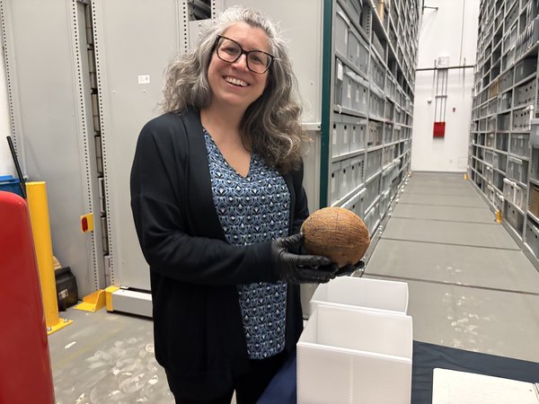
Nobel Prize-winning scientist Paul Lauterbur used a coconut to create the ground-breaking picture, which helped pave the way for MRI technology as we know it. That palm fruit, procured in the 1970s, is preserved in Philadelphia at the Science History Institute.
- INSIDE THE ARCHIVES
- PhillyVoice peeks into the collections at different museums in the city, highlighting unique and significant items you won't typically find on display.
How did a tropical treat get us to a medical tool that detects tumors, brain injuries and other life-threatening conditions? And why did it land in Old City roughly 50 years later? The story is just the kind of tale the museum and library, founded in 1982 to preserve and share histories of scientific discovery, loves to tell.
Early MRI experiments
Though scanning a coconut in a lab feels a bit primitive in 2025, it was a massive leap forward for science. MRI is an application of nuclear magnetic resonance, a phenomenon that occurs when a magnet is applied to atomic matter. The magnet affects the nuclei in a way that produces radio signals. Scientists already knew by the 1940s that analyzing these signals could help them understand the inner structure of whatever they were studying. They had developed NMR spectrometers to conduct these tests through the '50s and '60s. But they were limited and notoriously finicky. The Varian A-60, the first commercial model designed for routine use, came to the Science History Institute with a tongue-in-cheek "warning" printed next to the controls.
"A special circuit in the machine called a ‘CRISIS DETECTOR’ senses the operator’s emotional state in terms of how desperate he or she is to use the machine," the text read. "The ‘CRISIS DETECTOR’ then creates a malfunction proportional to the desperation of the operator. Threatening the machine with violence only aggravates the situation. Likewise, attempts to use any other machine may cause it to malfunction too — they belong to the same union."
- MORE INSIDE THE ARCHIVES
- Lewis and Clark's 200-year-old botanical treasures are preserved in Philadelphia
- How the Philadelphia Museum of Art unearthed forgotten anti-Nazi posters
- Sculptor who was the first Black woman to receive a national art commission was 'a voice of her time'
Still, the machine made it possible for more scientists to experiment with the technology. Lauterbur was one of them. The chemist had access to a Varian A-60 at the State University of New York at Stony Brook, where he was an associate professor. (He'd actually helped write the grant that allowed the college to purchase one.) Intrigued, he began visiting the lab after his usual hours.
"He wasn't really supposed to be doing this and he didn't want his secret out," said Michelle DiMeo, vice president of collections and programs for the Science History Institute.
Lauterbur also had a side gig in the summer of 1971 as the chairman of a failing Pennsylvania company specializing in NMR technology and headquartered just outside Pittsburgh. He was there when he got his big idea, apparently mid-meal. As Lauterbur often told it, he was grabbing a burger when inspiration struck. He wrote out his basic idea for an experiment that would create "a three-dimensional map of the distribution of particular classes of nuclei... within a living organism" in a composition notebook, dating and signing each of the six pages.
"We have a lot of archives with scientists racing to determine something," DiMeo said. "So it's important to lay your claim and have your dates."
Using this spectrometer on humans was out of the question, since the consoles were far too small to accommodate a person. A coconut, however, was the perfect candidate.
Taking the picture
Lauterbur didn't just choose a coconut because it fit inside his machine. Part of the attraction was that it was inanimate, so there was little risk of it squirming through the scans. Though the coconut was a relatively simple subject, its shell, inner lining and pulp also provided different materials for Lauterbur to analyze.
After producing two sets of 16 two-dimensional images, he was able to create a "true" three-dimensional graphic of the fruit. It was later published in a scientific journal in 1981.
The coconut gave way to more complex test subjects. Lauterbur worked his way from pine branches, nuts and clams up to live mice and human organs. He didn't work on a machine capable of imaging humans until he acquired "Big Red," a brightly painted contraption bursting with barely concealed wires. Its circular opening was actually too small for most people, so he had to wait for the manufacturer to deliver an updated model, "Big Red 2," in 1977.
Competing claims
At the same time that Lauterbur was installing "Big Red 2" in his Stony Brook lab, another scientist was making his own moves. Raymond Damadian, a Brooklyn-based medical doctor and research, scanned one of his assistants with his own machine in 1977. His company, Fonar Corporation, would produce the first commercial MRI scanner in 1980.
Damadian, and by extension Fonar, long claimed to be the inventor of magnetic resonance scanning. He chafed at competitors whom he claimed stole his proprietary technology, suing several manufacturers over copyright infringement — and winning $129 million from General Electric. When Lauterbur and the English physicist Sir Peter Mansfield received the Nobel Prize in medicine in 2003 for their discoveries in magnetic resonance imaging, Damadian protested his exclusion quite publicly.
In an ad that ran in several major newspapers, Damadian called his snub "the shameful wrong that must be righted" and provided testimonials from sympathetic colleagues. He encouraged readers to clip out and mail a coupon at the bottom addressed to the Nobel Prize Committee demanding his inclusion.
Lauterbur was, at the very least, inspired by Damadian's research into NMR. He credited a colleague's reproduction of a Damadian study on cancerous rat tissues with spurring his inquiry into non-invasive NMR imagery.
Preserving eureka moments
The Science History Institute acquired Lauterbur's collection from his widow after the scientist passed in 2007. It consists of roughly 50 boxes of papers, slides and objects like the coconut and notebook where he recorded his breakthrough idea. The bulk of it is stored in a six-story archival building next to the museum. Standing inside it, it's hard not to think of the cavernous warehouse from "Raiders of the Lost Ark." Staffers have to use a cherry picker to grab many of the materials.
"The archival staff gets OSHA-certified (every three years)," DiMeo said. "Which is not a thing you learn in library school."
Along with Lauterbur's effects, the museum houses the collections of seven other Nobel Laureates. It also stewards a handwritten Isaac Newton manuscript, correspondence from J. Robert Oppenheimer, vintage medical advertisements, numerous instruments and an original Teddy Ruxpin animatronic toy. Some of the heavier items, like the 1,250-pound magnet component of the Varian A-60, are housed at an off-site facility in New Jersey.
Visitors to the museum won't see most of its roughly 7,000-piece collection. Only about 6% of it is on the floor at any given moment, and the Science History Institute is still finishing up its $3.3 million renovations, which temporarily closed the building for about four months. (The revamped lobby is expected to debut by May, while a gift shop and Hall of Minerals should be completed by September.) But they can browse the collection digitally. The museum has made the images of its cataloged objects available for free and open use, a policy DiMeo is "really proud of."
By making these scientific histories more accessible and telling the complete story, from notebook to coconut, the Science History Institute hopes to inspire future pioneers.
"You know what an MRI is," she said. "Maybe you've had one or someone you love had one, but you don't know the story behind it. And the story I think is actually a really fun one, of experimenting with things that maybe seem forbidden at the beginning. It's a story of creativity and it's a story of trying to help people and getting some of the work that was happening in the lab to be extracted from an analytical chemistry setting to something that has practical benefits for people."
Follow Kristin & PhillyVoice on Twitter: @kristin_hunt
| @thePhillyVoice
Like us on Facebook: PhillyVoice
Have a news tip? Let us know.