


August 07, 2019
 Source/Penn Medicine
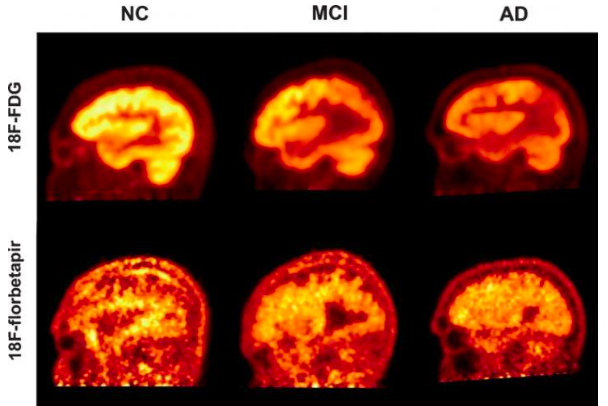
The University of Pennsylvania and Thomas Jefferson University teamed up on a study testing diagnostic methods to assess Alzheimer's disease. PET imaging shown above depict scans using the radiotracers FDG and florbetapir to quantify cognitive decline in patients with Alzheimer's disease, mild cognitive impairment and healthy controls.
A characteristic feature of Alzheimer's disease may not be the most accurate measure of its severity and progression in patients, according to new research from the University of Pennsylvania and Thomas Jefferson University.
The presence of amyloid plaque in the brains of Alzheimer's patients is a hallmark of the disease, but these protein deposits do not always correspond to the degree of cognitive decline patients are experiencing. The brains of those with mild cognitive impairment (MCI) or even no neurodegenerative symptoms may also show higher accumulations of amyloid.
In the new study out of Philadelphia, researchers offer evidence that measuring the brain's glucose consumption for neural activity is a more effective means of assessing cognitive impairment. They advocate for the use of fluorodeoxyglucose (FDG) PET scans over florbetapir-PET, which measure for amyloid protein.
"Both florbetapir-PET and FDG-PET are approved diagnostic methods for Alzheimer's disease, and both appear to be effective in indicating some sort of cognitive impairment," said the study's co-principal investigator Abass Alavi, a professor of radiology at Penn. "However, we have now shown that FDG-PET is significantly more precise in clinical studies, and it is also available for routine use with modest costs. Our results support the notion that amyloid imaging does not reflect levels of brain function, and therefore it may be of limited value for assessing patients with cognitive decline."
Researchers evaluated 63 individuals using both diagnostic tests. The study participants included 19 with clinically diagnosed Alzheimer's disease, 23 with MCI and 21 healthy individuals. All participants were assessed with a Mini Mental Status Examination (MMSE), a common diagnostic test used in the current study to gauge impairment and map the results to different regions of the brain based on the scans.
While both scan types were able to distinguish between the healthy group and individuals with dementia, the correlation between FDG and low cognitive performance across all groups in the study was more accurate than florbetapir-PET.
"Amyloid imaging has a value in diagnosing or ruling out Alzheimer's disease, but it's a bit like all or nothing. Our study shows that it can reveal disease, but you wouldn't be able to differentiate between someone who had very mild or very severe symptoms," said co-principal investigator Andrew Newberg, a professor of radiology at Thomas Jefferson University.
The results of the study are detailed in the August issue of the Journal of Alzheimer's Disease.