


January 12, 2015
 Thom Carroll/PhillyVoice
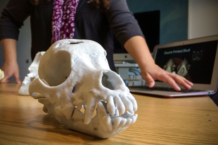
Dr. Evelyn M. Galban of the University of Pennsylvania School of Veterinary Medicine explains how 3-D prints are made by using images produced through CT scans.
PHOTOS: Penn Vet innovates using 3D printers
Even if these pictures are composited together on-screen to make a 3-D image, surgeons may enter a procedure without knowing exactly what a mass or malformation looks like as a whole until after they open up the patient. By inspecting a detailed 3-D printed model, this uncertainty can be diminished.
“The actual print allows an exact 'feel' and exact understanding of the problem in multiple planes,” said veterinary surgeon Dr. Ron Ben-Amotz at the Veterinary Specialty and Emergency Center (VSEC) in Philadelphia and Bucks County.
Take the case of a 10-year-old male black Labrador whose owner brought him in to Penn Vet's Ryan Hospital when he started to have seizures. Seizures in dogs are the most common indication of a brain tumor, particularly in those over 5 years old. The team of veterinarians — consisting of neurosurgeons, radiologists and radiation-oncologists — performed an MRI and immediately saw a large white mass crowding his skull where his brain would ordinarily be.
Galban thought this patient would be an excellent candidate for pre-op 3-D printing so the surgeons could practice removing the unwieldy tumor on a to-scale replica.
“We basically Googled who had a 3-D printer, and PennDesign came up first because they are right on campus,” she said. “They were so helpful in printing the data into something useable.”
Stephen Smeltzer and Dennis Pierattini oversee the woodworking, metalworking and digital fabrication that go on at PennDesign's Fabrication Lab, a multi-room maker shop on campus for students to bring their digital designs to life. The two are used to creating abstract architectural elements, buildings and cityscapes — but Galban's dog skull was a first. It didn't take much for them to lend a helping hand.
“We felt like we were actually helping the animals,” said Smeltzer. “We went over there and met with people from the staff, and there really was a dialogue between the two of us.”
The lab acquired its two 3-D printers about a decade ago. The main printer used by the Penn Vet team uses gypsum powder, since the final product's consistency is closer to that of bone. Special software transforms the MRI images into a printable format, and the data is sent to the printer. Models are built up layer-by-layer in an additive fashion, with an inkjet print head spraying certain sections of each layer with a liquid binding material to solidify the powder.
The excess gypsum is removed from non-bound areas, like eyeholes and areas between teeth, and the final model is sealed with a superglue-like substance for durability. The whole process can be completed overnight.
The end result is a finely detailed, solid white skull that looks and feels close to the real thing.
In the Labrador's case, Smeltzer printed not only the entire skull but also the cancerous mass inside the cranial cavity. While practicing the tricky procedure on the model, the team discovered the best way to approach the tumor: Cut a diamond-shaped piece of bone off the top of the cranium and go through the sinus into the braincase.
“It's unbelievably difficult to go off of an MRI only to know where your tumor is, so we then knew our approach would be appropriate,” Galban said.
The actual surgery was a resounding success. Her patient is healthy again, with no tumor recurrence thus far, to the immense relief of the owner.
“It's easier to talk about it with owners and show them,” Galban said. “You can be describing what you're seeing — a skull fracture, for instance — but a 3-D model makes it really easy to say, 'Look, here's the problem, and this is what we're doing to fix it.'”
The technology of 3-D printing, also known as additive manufacturing, is experiencing a boom of interest from just about every industry under the sun. In the 1980s, it served as a way for engineers and industrial designers to create rapid prototypes and experiment with new products.
Today, medical and dental professionals are using the technology for patient-customized implants and models. In the fashion and craft worlds, designers are showing off their 3-D-printed clothes, jewelry and costume pieces. There are even 3-D-printed foods like pizza, chocolate and cookies.
A handful of other veterinary institutions are printing pre-op 3-D models of their patients as well, such as Cornell University's College of Veterinary Medicine and Auburn University’s Veterinary Teaching Hospital.
Although VSEC doesn't have its own 3-D printer, Ben-Amotz sends CT data to his colleagues at Ohio State University, where he completed his residency. As an expert in wound management and fracture repair, he mainly uses the technology for complex orthopedic cases.
“The advantage of such a printer is that it allows the surgeon to better plan for surgery and better understand the true 3-D picture,” he said. “I use it to understand bone deformities before planning for corrective orthopedic procedures.”
With four cases under her belt so far, Galban hopes to proceed with more 3-D models this year and expand to different neurosurgical areas such as spinal malformations. She foresees unlimited utility with the technology in a veterinary setting, in terms of vet training, implant design and surgical planning.
“I see it continuing to move forward, especially with the new generation of printers out there,” said Smeltzer. “There’s printers that can print flexible materials now, so you could put tendons in there, or other soft matter.”