


July 14, 2020
 Source/Radiological Society of North America
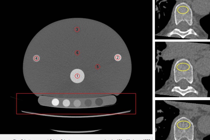
Using Mindways softwares, the above CT scan shows volume of interest (yellow circles) in the trabecular bone compartment of three vertebrae for bone mineral density measurements.
Bone mineral density tests are the current standard used to diagnose osteoporosis, a disease that weakens the bones and increases the risk of fracture.
Still, health experts say many at-risk people are not getting evaluated. But adding a bone mineral density test to a cardiac CT exam could improve the rate of detection, a new study finds.
Some 200 million people worldwide suffer from osteoporosis. While there are drugs that can be prescribed to reduce the risk of fractures, early detection is crucial. Almost 20% of Medicare fee-for-service beneficiaries in the U.S. died within 12 months of a new osteoporotic fracture, according to a National Osteoporosis Foundation report.
"Osteoporosis is a prevalent, under-diagnosed and treatable disease associated with increased morbidity and mortality," said lead study author Dr. Josephine Therkildsen, of Herning Hospital in Denmark. "Effective anti-osteoporotic treatment exists and so identifying individuals with greater fracture rate who may benefit from such treatment is imperative."
While the cardiac CT was designed to assess heart health, the scan also can visualize the thoracic vertebrae, the part of the spine found in the upper back. And it is easy to add a BMD test to it, the researchers said.
Doing so won't increase exam time or radiation exposure. Non-enhanced CT images work well as long as the correct calibration system and imaging acquisition techniques are used, Therkildsen said.
During the study, 1,487 participants underwent cardiac CT for evaluation of heart disease combined with BMD testing of three thoracic vertebrae using quantitative CT software.
Twelve percent of them had low BMD and 5.3% were diagnosed with a fracture within three years. The fracture was caused by osteoporosis in 31 of the 80 patients.
"We believe that opportunistic BMD testing using routine CT scans can be done with little change to normal clinical practice and with the benefit of identifying individuals with a greater fracture rate," Therkildsen said.
In theory, any routine CT image that includes a view of the thoracic vertebrae could work, researchers said. More research is needed to confirm the BMD cut-off values for treatment and provide more data on fracture risk based on gender and area of the body. Clinical risk factors also need to be considered.
Developing a fully automated software for BMD measurements would make this approach even more effective, researchers said.
"Our research group is dedicated to extend the research in this field, as we believe it should be added to clinical practice," Therkildsen said.
The study was published in Radiology.